


Diagnostic imaging provides medical professionals with a clear view of internal bodily structures. One of the most common imaging methods utilized in gynecology and obstetrics relies on sound waves, rather than radiation. The ultrasound gives doctors the ability to observe pregnancies and evaluate organ function. Understanding the purpose, frequency, and mechanics of this procedure helps patients prepare for their appointments and participate actively in their own healthcare decisions.
What Is an Ultrasound?
Medical providers utilize specific equipment to generate pictures of the inside of the human body. An ultrasound relies on high-frequency sound waves and operates by sending these sound pulses through the skin and into the internal structures. As the sound waves reach different organs, tissues, and fluids, they bounce back toward the source.
The resulting visual representations can be viewed as still photographs or moving video sequences on a monitor. Because the procedure utilizes sound waves instead of radiation, it serves as a routine option for examining delicate tissues and monitoring fetal development. The data collected from these images gives medical professionals a reliable method to identify structural anomalies and track biological changes over time.
When Should You Have One?
Doctors recommend this imaging procedure for several distinct reasons, which range from routine pregnancy monitoring to investigating unexplained physical symptoms. If a patient experiences heavy menstrual bleeding or severe pelvic pain, a provider might order a scan to examine the uterus and ovaries. The images can reveal physical signs of conditions like uterine fibroids. By observing the pelvic organs directly, the medical team may formulate an appropriate treatment plan tailored to the specific findings.
During pregnancy, this imaging tool tracks the growth and development of the fetus, the placenta, and the amniotic fluid. Patients typically undergo two scheduled scans, and the initial appointment can take place early in the first trimester. This early evaluation confirms the pregnancy, identifies the number of embryos, and helps the provider calculate the estimated gestational age. A second routine scan generally occurs around the 20-week mark.
What Does the Process Involve?
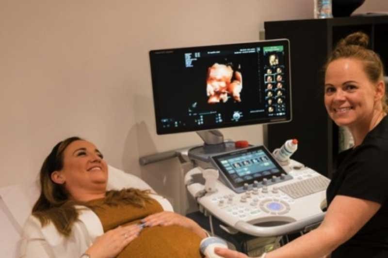
This diagnostic evaluation is a noninvasive and painless procedure that requires minimal physical preparation from the patient. During the appointment, the individual lies on an examination table and adjusts their clothing to expose the abdominal area. The technician begins by applying a specialized water-based gel directly to the skin. This substance facilitates the secure transmission of sound waves. The technician glides a handheld instrument, called a transducer, across the gel-covered skin. The transducer emits the sound pulses and records the echoes as they return from the internal organs.
As the transducer moves, the corresponding images appear on the nearby computer screen in real-time. The operator may pause to capture specific measurements or take still pictures for the medical file. Once the necessary views are obtained, the patient simply wipes off the gel and redresses. The medical provider then reviews the collected images to formulate a diagnosis or provide a detailed update on fetal development.
Seek Professional Women’s Health Care
Maintaining regular communication with a medical provider helps individuals navigate their reproductive health effectively. After completing an ultrasound, the doctor will review the findings and discuss any necessary next steps based on the visual evidence. Patients should always communicate openly about their physical symptoms and medical history. Establishing a relationship with a trusted gynecologist or obstetrician allows for more accurate diagnoses and personalized care plans. Whether you need a routine prenatal checkup or an evaluation for sudden pelvic discomfort, consulting an expert provides the guidance needed to protect your well-being. Routine assessments empower individuals to make educated choices regarding their physical health and medical treatments.


Leave a Reply