


Skin cancer affects millions of people every year, and among the most frequent diagnoses is squamous cell carcinoma. Left untreated, these cellular growths can spread and cause health complications. Medical professionals utilize specialized techniques to address these formations. One highly effective method is Mohs surgery. This procedure targets cancerous cells while preserving surrounding healthy tissue.
What Is Squamous Cell Carcinoma?
The skin serves as the body’s primary protective barrier against environmental elements, and the epidermis acts as the outermost shield in this system. Squamous cell carcinoma represents approximately two out of every ten skin cancer cases and originates directly in this top layer. Within the epidermis, flat squamous cells continuously shed as new ones form. When these specific cells begin to multiply uncontrollably, they create cancerous growths.
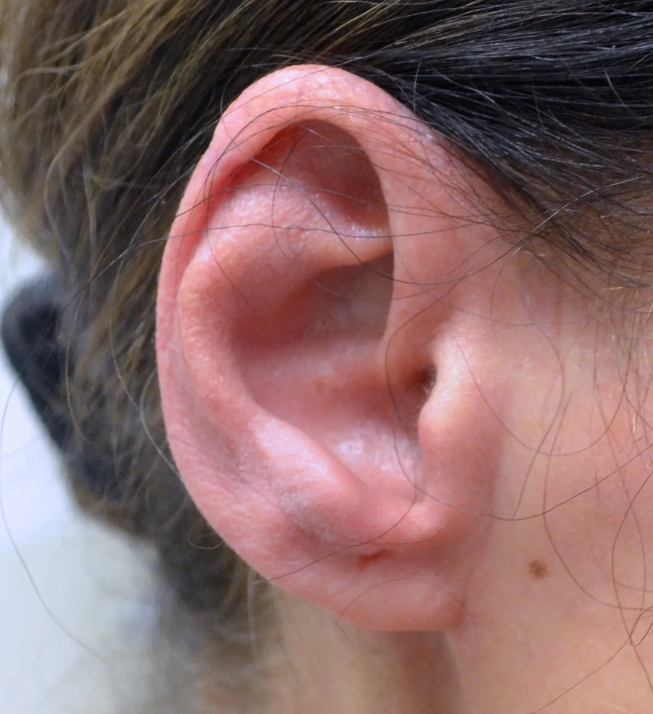
Patients typically notice early signs through physical changes on their skin. The condition frequently presents as reddish, scaly, or crusted patches. These formations require ultraviolet light exposure to develop, so they usually emerge on the face, neck, and the backs of the hands. Recognizing these early visual indicators prompts many individuals to seek a professional medical evaluation. Since the disease manifests on the skin’s surface, physicians can frequently detect it in its preliminary stages.
How Is It Detected and Diagnosed?
Medical professionals follow a structured process to evaluate suspected skin cancers. A specialist inspects the patient’s body from head to toe, looking for unusual spots, non-healing sores, or irregular patches. If a physician identifies a suspicious lesion, they will perform a biopsy. This procedure requires numbing the site with a local anesthetic before surgically removing a portion of or the entire growth. The extracted tissue then undergoes a rigorous evaluation under a high-powered microscope, as this laboratory analysis provides the only definitive method to confirm the presence of squamous cell carcinoma. Once the laboratory results verify the diagnosis, the medical provider discusses appropriate treatment plans with the patient.
How Does Mohs Surgery Work?
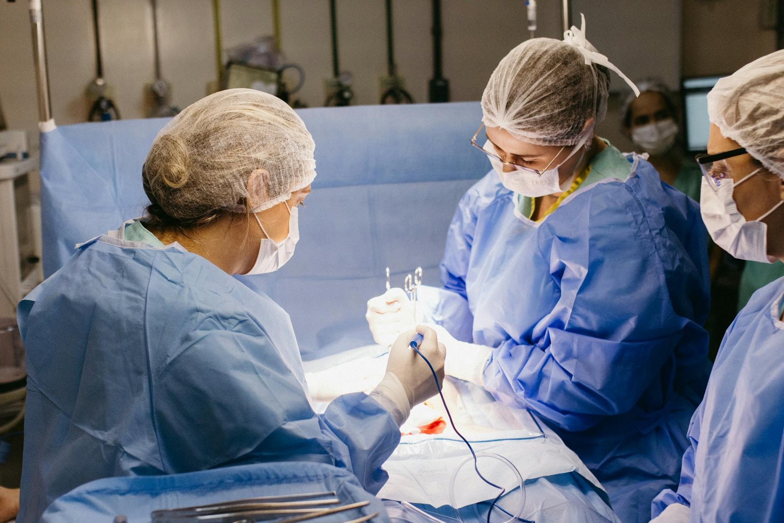
Specialists utilize Mohs surgery as a targeted approach for removing skin cancer. The surgeon begins by administering a local anesthetic to numb the target area. Precise surgical instruments are used to remove the visible portion of the tumor. The removed tissue is prepared for immediate microscopic examination, and the surgeon evaluates the sample to identify any remaining cancer cells along the margins. If malignant cells persist, the provider removes another targeted layer of tissue solely from the exact location where the cancer remains.
This cyclical process of removal and examination continues until the tissue samples show no signs of disease. By evaluating the entirety of the surgical margins, the Mohs technique effectively eliminates the carcinoma while preserving healthy surrounding tissue. The physical removal of a tissue layer takes only ten to fifteen minutes, but the laboratory preparation requires at least an hour. Patients should plan to dedicate a significant portion of their day to the appointment. Following the complete removal of the cancer, the surgeon determines the optimal method for closing the wound, which may involve natural healing, stitches, or a skin graft.
Meet With a Mohs Surgeon
Consulting a qualified medical professional provides the best path forward for addressing skin abnormalities. A dermatologist with specialized training in Mohs surgery possesses the specific skill set required to manage squamous cell carcinoma effectively. Post-operative care may require limiting strenuous physical activity and adhering to strict wound-care instructions to promote proper healing. Scheduling a routine skin cancer surveillance exam establishes a baseline for your skin health, allowing a specialist to monitor any changes over time.

Leave a Reply